Latest News & Promos
- Details
- Written by The Community Eyecare Team
A wrinkle on the retina - which is also known as an epiretinal membrane (ERM) or a macular pucker - is a thin, translucent tissue that develops on the surface of the retina.
The retina is the inner layer that lines the inside of the back of the eye and is responsible for converting the light image into an electrical impulse that is then transmitted to the brain. An epiretinal membrane that forms on the retina goes unnoticed by the patient many times, and is only noticed during a dilated eye exam by an eye doctor.
Epiretinal membranes can become problematic if they are overlying the macula, which is the part of the retina that is used for sharp central vision. When they become problematic they can cause distortion of your vision, causing objects that are normally straight to look wavy or crooked.
Causes of a wrinkle on the retina
The most common cause is age-related due to a posterior vitreous detachment, which is the separation of the vitreous gel from the retina. The vitreous gel is what gives the eye its shape, and it occupies the space between the lens and the retina. When the vitreous gel separates from the retina, this can release cells onto the retina surface, which can grow and form a membrane on the macula, leading to an epiretinal membrane.
ERMs can also be associated with prior retinal tears or detachments, prior eye trauma or eye inflammation. These processes can also release cells onto the retina, causing a membrane to form.
Risk factors
Risk for ERMs increases with age, and males and females are equally affected.
Both eyes have ERMs in 10-20% of cases.
Diagnostic testing
Most ERMs can be detected on a routine dilated eye exam.
An optical coherence tomography (OCT) is a noninvasive test that takes a picture of the back of the eye. It can detect and monitor the progression of the ERM over time.
Treatment and prognosis
Since most ERMs are asymptomatic, no treatment is necessary. However, if there is significant visual distortion from the ERM or significant progression of the membrane over time, then surgical intervention is recommended. There are no eye drops, medications, or nutritional supplements to treat or reverse an ERM.
The surgery is called a vitrectomy with membrane peeling. The vitrectomy removes the vitreous gel and replaces it with a saline solution. The epiretinal membrane is then peeled off the surface of the retina with forceps.
Surgery has a good success rate and patients in general have less distortion after surgery.
Article contributed by Dr. Jane Pan
This blog provides general information and discussion about eye health and related subjects. The words and other content provided in this blog, and in any linked materials, are not intended and should not be construed as medical advice. If the reader or any other person has a medical concern, he or she should consult with an appropriately licensed physician. The content of this blog cannot be reproduced or duplicated without the express written consent of Eye IQ.
- Details
- Written by The Community Eyecare Team
It could be a retinal vein occlusion, an ocular disorder that can occur in older people where the blood vessels to the retina are blocked.
The retina is the back part of the eye where light focuses and transmits images to the brain. Blockage of the veins in the retina can cause sudden vision loss. The severity of vision loss depends on where the blockage is located.
Blockage at smaller branches in the retinal vein is referred to as branch retinal vein occlusion (BRVO). Vision loss in BRVO is usually less severe, and sometimes just parts of the vision is blurry. Blockage at the main retinal vein of the eye is referred to as central retinal vein occlusion (CRVO) and results in more serious vision loss.
Sometimes blockage of the retinal veins can lead to abnormal new blood vessels developing on the surface of the iris (the colored part of your eye) or the retina. This is a late complication of retinal vein blockage and can occur months after blockage has occurred. These new vessels are harmful and can result in high eye pressure (glaucoma), and bleeding inside the eye.
What are the symptoms of a retinal vein occlusion?
Symptoms can range from painless sudden visual loss to no visual complaints. Sudden visual loss usually occurs in CRVO. In BRVO, vision loss is usually mild or the person can be asymptomatic. If new blood vessels develop on the iris, then the eye can become red and painful. If these new vessels grow on the retina, it can result in bleeding inside the eye, causing decreased vision and floaters – spots in your vision that appear to be floating.
Causes of retinal vein occlusion
Hardening of the blood vessels as you age is what predisposes people to retinal vein occlusion. So retinal vein occlusion is more common in people over the age of 65. People with diabetes, high blood pressure, blood-clotting disorders, and glaucoma are also at higher risk for a retinal vein occlusion.
How is retinal vein occlusion diagnosed?
A dilated eye exam will reveal blood in the retina. A fluorescein angiogram is a diagnostic photographic test in which a colored dye is injected into your arm and a series of photographs are taken of the eye to determine if there is fluid leakage or abnormal blood vessel growth associated with the vein occlusion. An ultrasound or optical coherence tomography (OCT) is a photo taken of the retina to detect any fluid in the retina.
Treatment for retinal vein occlusion
Not all cases of retinal vein occlusion need to be treated. Mild cases can be observed. If there is blurry vision due to fluid in the retina, then your ophthalmologist may treat your eye with a laser or eye injections. If new abnormal blood vessels develop, laser treatment is performed to cause regression of these vessels and prevent bleeding inside the eye. If there is already a significant amount of blood inside the eye, then surgery may be needed to remove the blood.
Outlook after retinal vein occlusion
Prognosis depends on the severity of the vein occlusion. Usually BRVO has less vision loss compared to CRVO. The initial presenting vision is usually a good indicator of future vision. Once diagnosed with a retinal vein occlusion, it is important to keep follow-up appointments to ensure that prompt treatment can be administered to best optimize your visual potential.
Article contributed by Dr. Jane Pan
This blog provides general information and discussion about eye health and related subjects. The words and other content provided in this blog, and in any linked materials, are not intended and should not be construed as medical advice. If the reader or any other person has a medical concern, he or she should consult with an appropriately licensed physician. The content of this blog cannot be reproduced or duplicated without the express written consent of Eye IQ.
Patient Resources
Please Leave a Google Review
We sincerely appreciate our patients and welcome your feedback. Please take a moment to let us know how your experience has been.
Our Dry Eye Center
Northeast Wisconsin's Optometrists of Choice
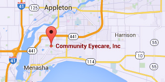
Menasha Office
1255 Appleton Road
Menasha, WI 54952-1501
Phone: (920) 722-6872
CALL 1ST - NO WALK-INS
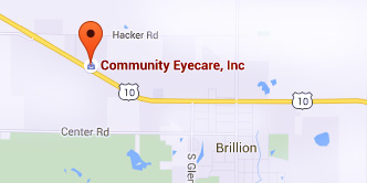
Brillion Office
950 West Ryan Street
Brillion, WI 54110-1042
Phone: (920) 756-2020
CALL 1ST - NO WALK-INS
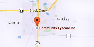
Black Creek Office
413 South Main Street
Black Creek, WI 54106-9501
Phone: (920) 984-3937
CALL 1ST - NO WALK-INS
© Community Eyecare, Inc.: 3 Convenient Northeast Wisconsin Locations | Menasha, WI | Brillion, WI | Black Creek, WI | Site Map
Text and photos provided are the property of EyeMotion and cannot be duplicated or moved.


